படிமம்:Paramecium.jpg
Jump to navigation
Jump to search

இந்த முன்னோட்டத்தின் அளவு: 228 × 209 படப்புள்ளிகள் . மற்ற பிரிதிறன்: 751 × 689 படப்புள்ளிகள் .
மூலக்கோப்பு (751 × 689 படவணுக்கள், கோப்பின் அளவு: 183 KB, MIME வகை: image/jpeg)
சுருக்கம்
| விளக்கம் |
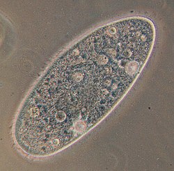
Deutsch: Paramecium aurelia - Optisches Mikroskop. Paramecium aurelia, der bekannteste von allen ciliaten. Die Blasen innerhalb der Zelle sind Vakuolen. Die gesamte Oberfläche ist mit Wimpern umgeben, die durch ihre schnelle Bewegung verwischt werden.
English: Paramecium aurelia. Optical microscope. Paramecium aurelia, the best known of all ciliates. The bubbles throughout the cell are vacuoles. The entire surface is covered in cilia, which are blurred by their rapid movement.
Français : Paramecium aurelia. Microscope optique. Le plus connu des ciliés. Les bulles que vous voyez sont des vacuoles. Tout le corps est couvert par des cils, qui sont flous sur l'image à cause de leurs mouvements rapides.
Polski: Paramecium aurelia - pantofelek, najbardziej znany ze wszystkich orzęsków. Bąbelki w środku komórki to wodniczki. Cała powierzchnia pantofelka pokryta jest rzęskami, które są na fotografii zamazane ze względu na ich szybki ruch.
Српски / srpski: Paramecium aurelia, najpoznatiji od svih trepljara pod optičkim mikroskopom. "Mehurići" u ćeliji paramecijuma su vakuole. Cela površina tela je prekrivena trepljama, koje su na slici mutne zbog toga što se brzo pokreću.
Türkçe: Paramecium aurelia - optik mikroskop. Paramecium aurelia, tüm siliyalılar içinde en çok bilinen türdür. Hücre boyunca yuvarlak olarak izlenen oluşumlar, vakuollerdir. Hücrenin tüm yüzeyi, hızlı hareketlerinden dolayı bulanık görüntü vermiş olan siliya ile kaplıdır. |
| நாள் | |
| மூலம் | Originally uploaded to the English Wikipedia, where it was made by Barfooz. |
| ஆசிரியர் | Barfooz at the English Wikipedia. |
| ஒத்தக்கோப்பு | Transparent |
அனுமதி

|
GNU Free Documentation License விதிமுறைகளின் கீழ் இந்த ஆவணத்தை நகலெடுக்க, விநியோகிக்க மற்றும்/அல்லது மாற்றுவதற்கு அனுமதி வழங்கப்பட்டுள்ளது, Free Software Foundation;ஆல் வெளியிடப்பட்ட பதிப்பு 1.2 அல்லது அதற்குப் பிந்தைய பதிப்பு, மாற்றமில்லாத பிரிவுகள், முன் அட்டை உரைகள் மற்றும் பின் அட்டை உரைகள் இல்லாமல் வெளியிடப்பட்டது. GNU Free Documentation License என்ற தலைப்பில் உரிமத்தின் நகல் சேர்க்கப்பட்டுள்ளது. |
| இந்த கோப்பு Creative Commons Attribution-Share Alike 3.0 Unported உரிமத்தின் கீழ் உள்ளது. | ||
| ||
| This licensing tag was added to this file as part of the GFDL licensing update. |
Soft scrubbed view
Original upload log
Originally uploaded to English Wikipedia.
- 23:11, 27 October 2004 . . Barfooz (Talk) . . 751x738 (190517 bytes) (Paramecium viewed under a microscope)
- 15:19, 28 June 2004 . . Josh Grosse (Talk) . . 236x152 (3913 bytes) (Reverted to earlier revision)
- 15:19, 28 June 2004 . . Josh Grosse (Talk) . . 236x152 (5129 bytes) (Reverted to earlier revision)
- 15:13, 28 June 2004 . . Josh Grosse (Talk) . . 236x152 (3913 bytes) (Better image, created by self)
- 20:04, 10 October 2003 . . Josh Grosse (Talk) . . 236x152 (5129 bytes)
கோப்பின் வரலாறு
குறித்த நேரத்தில் இருந்த படிமத்தைப் பார்க்க அந்நேரத்தின் மீது சொடுக்கவும்.
| நாள்/நேரம் | நகம் அளவு சிறுபடம் | அளவுகள் | பயனர் | கருத்து | |
|---|---|---|---|---|---|
| தற்போதைய | 05:58, 4 சூலை 2025 | | 751 × 689 (183 KB) | wikimediacommons>Chiswick Chap | Cropped 7 % vertically, 7 % areawise using CropTool with precise mode. |
கோப்பு பயன்பாடு
பின்வரும் பக்க இணைப்புகள் இப் படிமத்துக்கு இணைக்கபட்டுள்ளது(ளன):